
記住我


A randomized controlled trial was conducted in the First Affiliated Hospital of the University of Science and Technology of China (USTC). The study was approved by the ethics committee of the First Affiliated Hospital of USTC (approval no. KY2022-337). This trial was registered in the Chinese Registry of Clinical Trials (http://www.chictr.org.cn; registration no. ChiCTR2200066821) on 19/12/2022. Prior to the study, written informed consent was obtained from all participants.
We enrolled patients who were aged 18–75 years, had ASA physical status I-III, had SARS-CoV-2 infection 3 months prior to surgery, and had received elective thoracoscopic lung surgery using DLTs from December 2022 to April 2023. The following patients were excluded: (1) those who refused to participate; (2) those with a recent incidence of sore throat or upper respiratory tract infection orhoarseness; (3) those with a Mallampati score of 4; and (4) those with a history of difficult airway.
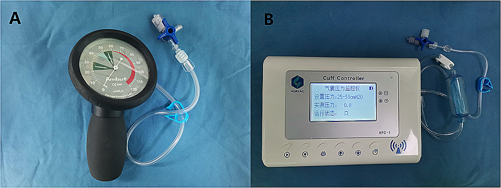
Each individual was randomly assigned to the thermal softening group or the control group in a 1:1 ratio on the basis of a random table generated by a computer randomization program. The random sequence was placed in opaque, sealed envelopes. Patients, anesthesiologists, surgical team members, nurses, and postoperative follow-up evaluators were blinded to group assignment. After patients entered the operating room, the envelope was opened by a researcher who was also blinded to group allocation. The researcher prepared and pretreated all DLTs(Tuoren Best Medical Equipment Company, Henan, China). The size of the DLT depended on the patients’ gender and height [12]. The researcher first evacuated the air of the tracheal cuff and bronchial cuff and then immersed the distal part of the DLTs in a sterile saline solution for 10 min (Fig. 1). The temperature of the saline was maintained at 50 °C for the thermal softening group or at room temperature for the control group; a thermostatic kettle was used to measure saline temperature. The temperature was regulated at the desired level, and following the completion of thermal softening, the DLTs were removed from the saline solution bottle before tracheal intubation.
Fig. 1
The thermal softening process for the DLT. (A) The distal portion of the DLT was placed in a sterile saline solution bottle, and the bottle was kept on a constant temperature kettle whose temperature could be adjusted. (B) The constant temperature kettle was covered with a layer of cloth to blind hospital staff regarding group allocation
Once the patient entered the operating room, blood oxygen saturation, electrocardiography, heart rate, noninvasive blood pressure, and bispectral index (BIS) were continuously monitored. Invasive arterial blood pressure was monitored by radial artery puncture and catheterization after local anesthesia.
Anesthesia was induced using midazolam (0.05 mg/kg), etomidate (0.3 mg/kg), sufentanil (0.5 µg/kg), and cisatracurium (0.2 mg/kg). An otorhinolaryngologist blinded to group allocation evaluated the vocal cords of the patients before tracheal intubation by using a video laryngoscope and acquired photographs for subsequent comparison. Tracheal intubation was performed by an anesthesiologist with experience in performing over 500 DLT insertions by using a video laryngoscope (INSIDES Medical Technology Company, Shenzhen, China). Endotracheal intubation was performed with a disposable polyvinyl chloride DLT by using a video laryngoscope. After inserting the tip of the bronchus forward through the glottis, the anesthesiologist removed the tube core and continue pushing forward.
If substantial airway resistance was encountered, the anesthesiologist rotated the DLT counterclockwise or clockwise by 90° and then pushed it forward. If severe resistance was still experienced, the anesthesiologist again rotated the DLT counterclockwise or clockwise by 90° and subsequently advanced the DLT [4]. The anesthesiologist adjusted the position of the patient’s head and used a fiberoptic bronchoscope(FOB)(UE Medical Company, Zhejiang, China) to evaluate and adjust the position of the DLT [13]. After the patient was placed in the lateral position, the position of the DLT was again confirmed using the FOB. The pressure in the tracheal and bronchial cuff was adjusted to less than 25 cmH2O and 44cmH2O, respectively, by using a cuff pressure monitoring device [14]. General anesthesia was maintained using propofol at 4–8 mgkg− 1 h− 1 and remifentanil at 0.1–0.3 µgkg− 1 min− 1. The infusion rate of propofol and remifentanil was adjusted to control the depth of anesthesia within a BIS of 40 to 60.
Thirty minutes before the completion of the surgery, the patients were intravenously administered 1 mg kg− 1 flurbiprofen axetil and 0.1 mg kg− 1oxycodone for postoperative pain management. After surgery, all patients were shifted to the postanesthesia care unit(PACU). The patients were treated with 0.05 mg kg− 1 neostigmine and 0.01 mg kg− 1 atropine to antagonize residual neuromuscular block. Patient-controlled intravenous analgesia(PCIA) infusion was initiated after extubation for all patients. The PCIA infusion comprised 1 µg/mL sufentanil and 1 mg/mL flurbiprofen in normal saline at a continuous infusion rate of 2 mL h− 1 with a 2-mL bolus and a lockout interval of 15 min.
Measurement of outcomesThe Mallampati grade was assessed preoperatively by ananesthesiologist blinded to the study details. The anesthesiologist used a video laryngoscope to acquire photographs and record the images of the vocal cords during endotracheal intubation; the laryngoscopy field of view was graded using the Cormack and Lehane scoring system. The percentage of glottis exposure was recorded in the range of 0–100%. The resistance encountered by the anesthesiologist during the insertion of DLTs was categorized into four levels: no resistance, mild resistance, moderate resistance, and severe resistance. The intubation time was defined as the time between the insertion of the laryngoscope into the patient’s mouth and the removal of the laryngoscope. The following hemodynamic parameters were measured: heart rate and mean arterial pressure before intubation and 2 min after intubation.
Trachea and vocal cord injuries related to DLT intubation were assessed by a blinded otorhinolaryngologist with more than 5 years of experience in using an FOB. Before removing the DLT, the otorhinolaryngologist inserted the FOB into the DLT and evaluated trachea and vocal cord injuries during extubation. Tracheal injury was assessed using the tracheal injury clinical scoring systembased on mucosal changes [15]. Vocal cord injury was assessed based on the following characteristics: (1) edema, swollen mucosa at the vocal folds; (2) erythema, redness of the mucosa with inflammatory swelling of the surrounding area; and (3) hematoma, bleeding of the vocal cord [16, 17].
POST was observed in the PACU at 1 h after the surgery and in the ward at 6 and 24 h after the surgery by a researcher blinded to group allocation. The severity of POST was assessed as follows: 0, no sore throat; 1, mild (less than that observed in common cold); 2, moderate (similar to that observed in common cold); and 3, severe (more than that observed in common cold) [18, 19]. The incidence and severity of hoarseness were also recorded. Hoarseness was assessed as follows: 0, no hoarseness; 1, mild (no hoarseness at the time of interview but was present previously); 2, moderate (perceived only by the patient); and 3, severe (recognizable at the time of interview) [18, 19].
Statistical analysisIn the study of Parket al. [20], 58% of the patients in the control group complained of sore throat at 6 h following DLT insertion. A 50% reduction in the incidence of POST was considered clinically significant. Therefore, 52 patients were required in each group, with an alpha level of 0.05 and astatistical power of 80%. A 10% drop-out rate was allowed; thus, 60 patients were required in each group.
SPSS version 25.0 software (IBM, Chicago, IL, USA) was used for all statistical analysis. For continuous data, the Kolmogorov–Smirnov test was used to determine data normality. Normally distributed data were reported as mean ± SD and evaluated using an independent Student’s t-test for equal variance. Non-normally distributed interval and ordinal data were expressed as median (interquartile range) and evaluated using Mann–Whiney U test. Categorical variables were expressed as absolute numbers (%) and analyzed by the χ2 test. For all analyses, P < 0.05 (two-tailed) was considered significant.
留言 (0)