
記住我


Thyroid eye disease (TED) is an autoimmune disorder characterized by inflammation and swelling of the extraocular muscles and orbital fat that develops in up to 40% of patients with Graves disease (GD).1 Thyroid eye disease can cause pain, diplopia, eyelid retraction, exophthalmos, and in worse cases reduced vision, leading to significant functional and psychological impairment.2 Women are disproportionately affected, with a reported female-to-male ratio of 4.2:1, reflecting the higher incidence of GD in females.3 Thyroid eye disease typically develops in close temporal association with GD as 85% of patients develop eye symptoms within 18 months before or after the onset of thyroid dysfunction.4 In up to 30% of the cases, TED can precede the hyperthyroidism.5
The exact pathogenesis of TED is not completely understood. Evidence indicates that binding of antibodies to thyrotropin receptors (TRAb) and insulin-like growth factor 1 receptors on fibroblasts is essential, leading to release of inflammatory mediators and recruitment of bone marrow–derived fibrocytes and lymphocytes (both T and B cells) to the orbit.6 Supported by ex-vivo studies, cytokines can stimulate production of extracellular matrix components, like glycosaminoglycans and hyaluronic acid, which are deposited in the affected orbital tissues.7 In addition, there is an increase in the number and volume of orbital adipocytes. Collectively, these processes culminate in the expansion of soft tissue within the orbital cavity, impeding venous outflow and exacerbating venous stasis that leads to further tissue volume expansion.
At present, there is no cure for TED, and the therapeutic approach is primarily focused on managing symptoms and reducing inflammation. According to the European Group of Graves’ Orbitopathy guidelines, first-line treatment entails the use of glucocorticosteroids.8 Glucocorticosteroid therapy involves a wide range of adverse effects, and approximately 20% to 25% of the patients are nonresponders.9
In recent years, several biological drugs have been introduced as immunosuppressive alternatives. These drugs more selectively target the inflammatory cascade, with rituximab acting as a CD20 receptor inhibitor on B-cells, tocilizumab as interleukin (IL)-6 receptor antagonist, and teprotumumab inhibiting insulin-like growth factor 1 receptors on orbital fibroblasts.10
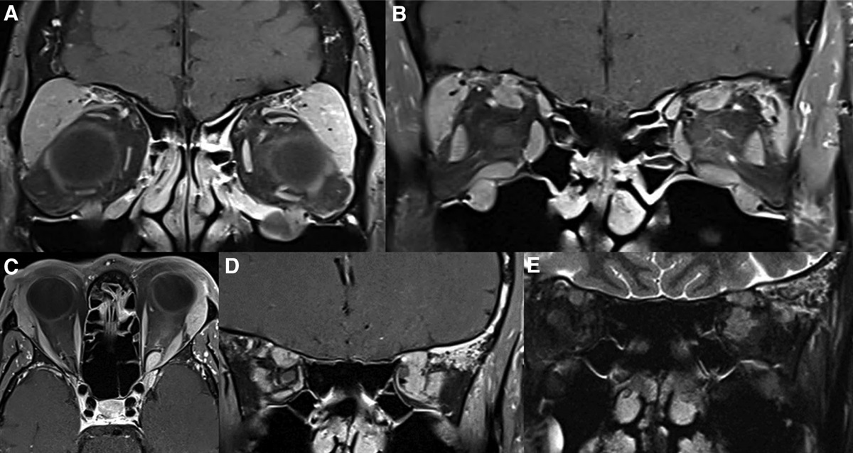
Diagnosis of GD relies on detection of serum TRAb.11 The same unambiguous diagnostic connection does not apply to TED, because many patients with elevated TRAb do not develop orbitopathy. The identification of TED patients is therefore mainly based on clinical findings. The Clinical Activity Score (CAS) measures inflammatory activity, and is a widely used assessment tool to guide treatment decision and monitor the effectiveness of therapy over time.12 Radiologic examination with CT or MRI can be helpful when there are only subtle signs of orbital inflammation (white-eyed-TED).13
A sensitive and specific biochemical marker for TED would be of great value, especially in subclinical cases, in patients with orbital manifestations preceding thyroid dysfunction, and in monitoring treatment effectiveness. A biomarker is also warranted to aid the assessment of GD patients selected for radioiodine treatment, as it is not possible to reliably predict in which patients TED will develop or worsen.14 In patients considered to be at risk of progression and/or development of TED during radioiodine treatment, European Group of Graves’ Orbitopathy guidelines recommend initiating oral prednisolone therapy, which should be gradually tapered off over a period of 3 months.8 In addition, identifying novel biomarkers is likely to shed light on the pathogenesis and provide new avenues for treatment.
To be applicable for broad clinical use, a biomarker should be easy to obtain without involvement of invasive procedures like biopsy. A wide range of molecules in blood, tears, and urine have been evaluated and proposed as potential biomarkers of TED. Most of these studies have been performed using traditional enzyme-linked immunosorbent assay, western blotting, and cell-based assays.15,16 Recent advances in proteomic techniques have enabled effective analysis of multiple proteins, providing the potential to identity novel molecular biomarkers. This article aims to present an overview of molecular biomarkers examined in TED.
Defining the Ideal TED Biomarker.The term biomarker has been defined in various ways. A frequently cited definition describes it as “a single indicator that objectively measures and evaluates normal or pathogenic biological processes.”17 The core function of a biomarker is to indicate the presence or severity of a particular disease or condition. Biomarkers can encompass any biologic substance, including proteins, nucleic acids, lipids, or other small molecules. In addition, specific clinical features, clinical scoring systems, and radiologic imaging may serve as biomarkers. A molecular biomarker can be detected in body fluids or tissue, such as blood, urine, tears, or fat.
Molecular biomarkers comprise genetic biomarkers (DNA mutations), protein biomarkers (antibodies, enzymes, hormones), and metabolic biomarkers (metabolites produced during metabolic processes).
For clinical utilization of a molecular biomarker in TED, the following criteria should be fulfilled:
Sensitivity: The biomarker should be able to detect the presence of the disease at an early stage. As such, it should accurately identify all or most patients having the disease. Specificity: The biomarker should be specific for the disease and not be affected by other conditions or factors. As such, it should be able to identify patients not having the disease. Reproducibility: The measurement of the biomarker should be consistent and accurate across laboratories commonly utilized in clinical settings. The biomarker must also demonstrate consistency when measured repeatedly under identical conditions. Easily accessible: A biomarker should be easy and safe to obtain, such as blood, tear, and urine samples. Clinical relevance: The biomarker should be useful for diagnosis, prognosis, or treatment. METHODSThis review focuses on studies evaluating the diagnostic performance of biomarkers in TED. A literature search was conducted on April 1, 2023, using PubMed and Embase. The following keywords were used: (“thyroid eye disease” OR “Graves’ orbitopathy” OR “Graves’ ophthalmopathy” OR “dysthyroid orbitopatopathy”) AND (“serum biomarker” OR “plasma biomarker” OR “blood biomarker” OR “tear biomarker” OR “urine biomarker” OR “biologic marker” OR “biological marker” OR “immune marker” OR “immunologic marker”).
A total of 242 articles were retrieved from this search. The articles were screened for eligibility against inclusion and exclusion criteria. Inclusion criteria were English-written original studies on molecular biomarkers for TED performed on blood, tear fluid, or urine samples. We excluded citations from letters, editorials, meeting reports, reviews, and case studies. Studies on biomarkers obtained from orbital and thyroid tissue were also excluded, since these require biopsy or surgery. Ultimately, 46 studies were included in this review (Fig. 1).
 FIG. 1.:
 PRISMA flowchart illustrating the identification, screening, and selection of studies. GD, Graves disease; PRISMA, Preferred Reporting Items for Systematic and Meta-Analysis; TED, thyroid eye disease.
This study did not require approval by an ethical committee as no original data were obtained.
RESULTS Validated Blood Biomarkers in Current Clinical Use AntibodiesAntibodies against thyroid-stimulating hormone (TSH) receptor (TSHR) were discovered over 60 years ago and are autoantibodies essential in the pathogenesis of GD.18 It binds to TSHR on the surface of thyroid cells most often with a stimulatory effect, leading to hyperthyroidism. However, they can also have a blocking or neutral effect in some cases. Anti-TSHR antibody is generally termed as TRAb and includes any subtype of antibody binding to the TSHR.19
Autoantibodies against TSHR can be quantified by traditional competitive-binding immunoassays, which measure the binding of antibodies to the TSHR.20 Such assays are commonly referred to as TSHR-binding inhibitory immunoglobulin (TBII) assays, or simply TBII assay. The principle behind TBII assay is that TRAb inhibits the binding of radiolabeled TSH to the TSHR, enabling quantitative measurement of total TRAb levels, regardless of function. The accuracy for TBII assays has improved with time and they are widely used in clinical practice.
High TRAb levels are associated with development of orbitopathy in patients with GD, and TRAb levels have been reported to correlate with inflammatory activity in TED.21–23 Patients with serum TRAb above 8.8 IU/L are at risk of a more severe disease course.24 Serial TRAb level measurements have been suggested as a guide for monitoring and treatment of TED.25
The TSHR stimulating immunoglobulin (TSI) is the stimulating subgroup of TRAb. The TSI can be measured in functional cell-based bioassays detecting the production of cyclic adenosine monophosphate produced in response to TSI binding of TSHR.19 The TSI levels therefore only reflect the stimulating part of TRAb. Cell-based bioassays are also capable of measuring TSHR blocking immunoglobulins (TBI). In current clinical practice, TSI and TBI bioassays are less available than TBII assays because they are more technically challenging to perform.
A multicenter study on 157 children with GD demonstrated that TSI bioassay is a more sensitive, specific, and reproducible technique than TBII assay.26 In this study, the level of TSI correlated with the incidence of TED. The superior sensitivity of TSI over TBII has later been confirmed in subcategories of TED, including euthyroid patients and mild cases of TED.27–29 There is evidence supporting a correlation between TSI levels and CAS, and some studies indicated that TSI levels also correlate with the severity of TED.30–32 Furthermore, TSI has been evaluated for predicting active TED with a sensitivity of 77.4%, specificity of 81.3%, and area under the curve (AUC) of 84.7%.33 Ponto et al.31 reported both high sensitivity (95%) and specificity (80%) using a TSI cut-off level of 377 specimen to reference ratio to identify TED patients with recent onset dysthyroid optic neuropathy (DON). Interestingly, Kahaly et al.21 demonstrated that a significantly higher dilution factor was needed to achieve undetectable TSI in GD patients with TED (1:6,561) compared with GD without TED (1:27). The authors proposed that this method could be clinically relevant for differentiating TED from non-TED.
Blood Biomarkers With Evaluated Diagnostic PerformanceStudies comparing biomarkers in GD patients with and without TED, and studies comparing patients with active and inactive TED, are particularly interesting for evaluating potential biomarkers in TED. Conversely, reported discrepancies in biomarker levels between TED and healthy controls (HC) may reflect changes associated with GD rather than orbitopathy, and hold less significance. Promising biomarkers evaluated in terms of sensitivity, specificity, and receiver operating characteristics are presented in the Table 1.
TABLE 1. - Studies reporting on diagnostic performance of biomarkers in TED Author (year) Sample size Assay Biomarkers Main findings Blood Cheng et al., 201834 TED n = 77ALDH2, aldehyde dehydrogenase 2; AUC, area under the curve; AZGP1, zinc-alpha-2 glycoprotein; CAS, Clinical Activity Score; CCL2, monocyte chemoattractant protein-1; CSF1, macrophage colony-stimulating factor 1; CYR61, cysteine-rich angiogenic inducer 61; DON, dysthyroid optic neuropathy; ELISA, enzyme-linked immunosorbent assay; FGF-21, fibroblast growth factor-21; Flt3L; Fms-related tyrosine kinase 3 ligand; GD, Graves disease (without orbitopathy); HC, healthy controls; IL, interleukin; LACRT, lacritin; LYZ, lysozyme C; PTX3, pentraxin-3; RhoA; Ras homolog family member A; TED, thyroid eye disease; TSI, thyroid-stimulating immunoglobulin.
Cysteine-rich angiogenic inducer 61 is a protein secreted by endothelial cells and fibroblasts. Cysteine-rich angiogenic inducer 61 is involved in a variety of cellular processes, including inflammation and cellular proliferation. Woo and coworkers41 observed that levels of cysteine-rich angiogenic inducer 61 were higher in TED than in HC, and higher in the active stage of TED compared with the inactive stage. There was no significant difference in cysteine-rich angiogenic inducer 61 between inactive TED and HC. They reported a sensitivity of 75.0% and specificity of 76.9% for cysteine-rich angiogenic inducer 61 as a diagnostic biomarker for active TED using a cut-off value of 116.5 pg/dL.
Ras homolog family member A is a protein that plays a role in adhesion and migration of T cells. A Chinese study proposed that elevated Ras homolog family member A levels in GD patients at baseline could be a biomarker for later development of TED with an AUC of 79.5%.43 In that study, the level of Ras homolog family member A was observed to be a more reliable predictor for TED than the level of TRAb.
Pentraxin-3 (PTX3) is a component of the innate immune system, and a member of the pentraxin family (which also includes c-reactive protein and serum amyloid P component). A study from 2018 reported elevated serum level of PTX3 in TED patients.38 The authors advocated that PTX3 could represent a diagnostic marker for TED with encouraging diagnostic performance (Table 1). However, in this study, the PTX3 levels were only compared between TED patients and HC, and there was no difference in PTX3 level between patients with active and inactive disease.
In a study from 2020 on various cytokines, elevation of both a Th1 chemokine (CXCL10) and Th17 cytokine (IL-23) in newly diagnosed GD patients was observed (Fig. 2). In the subgroup of GD patients with TED, they found elevation of a Th2 chemokine (CCL2) in combination with low selenium to be associated with high CAS.35 The authors reported a sensitivity of 83.3% and specificity of 85% for CCL2 levels above 190.5 pg/mL as an independent predictor for active TED. In addition, selenium levels below 64.3 µg/L were able to differentiate active from nonactive TED with a sensitivity of 88.9% and a specificity of 55%. This article did not evaluate the combination of CCL2 and selenium.
 FIG. 2.:
 The signature cytokines of Th-cell subsets. CXCL10, C-X-C Motif Chemokine Ligand 10; IFN-γ, interferon-γ; IL, interleukin; TGF-β, transforming growth factor beta; TNF-α, tumor necrosis factor-α. (This figure was generated using Servier Medical Art.)
A recent publication from Zhang and coworkers42 compared interleukins from the IL-12 family between TED patients and HC. The authors reported increased serum levels of IL-27 and IL-35 and suppressed levels of IL-12. As independent diagnostic markers to discriminate TED from HC, IL-27, IL-35, and IL-12 showed respective AUC values of 74%, 78%, and 78%.
Extracellular Matrix ProteinsFibulin-1 is an extracellular matrix protein secreted by orbital fibroblasts. It is involved in various cellular functions like adhesion, migration, and differentiation. Hu and coworkers36 reported higher plasma levels of fibulin-1 in GD patients with than without TED, in addition to higher fibulin-1 in active compared with in inactive TED patients. By using receiver operating characteristic analysis, fibulin-1 showed high sensitivity (93.3%) and specificity (88%) for predicting disease activity in TED at a cut-off value of 625.3 pg/mL.
Extraocular Muscle AntibodiesIn the early 2000s, there was a mounting interest in extraocular muscle antibodies in TED. Calsequestrin, a calcium-binding protein, is expressed 4-to-8 times higher in eye muscles than in skeletal muscles in other parts of the body. Antibodies directed toward calsequestrin were detected in 80% of patients with upper eyelid retraction, and later observed in 92% of patients with extraocular muscle involvement.46,47 One study observed that serum levels of calsequestrin antibodies were significantly higher in patients with active compared with inactive TED. The authors reported that the optimal cut-off level discriminating active from inactive TED was 138 ng/L with a sensitivity of 88.4%, specificity of 89.2% and AUC of 87.9%.39
Intracellular EnzymesAldehyde dehydrogenase 2 is a key enzyme in mitochondrial oxidation. Cheng and coworkers34 reported higher serum levels of anti–aldehyde dehydrogenase 2 in TED patients compared with HC. The authors defined a cut-off value of aldehyde dehydrogenase 2 as mean concentration in the serum of HC plus 3 standard deviations. Aldehyde dehydrogenase 2 then was able to identify active TED with a diagnostic sensitivity of 76.1% and a specificity of 78.6%. The authors did not present a receiver operating characteristic analysis in this study.
Growth FactorsOrbital volume expansion in TED is partly due to enhanced adipocytogenesis.48 Fibroblast growth factor-21 is known to stimulate glucose uptake in adipocytes via the induction of a glucose transporter. One study found that fibroblast growth factor-21 levels were higher in GD patients who later developed TED compared with those who did not.40 The authors reported that fibroblast growth factor-21 could predict the development of TED with a sensitivity 89%, a specificity 53%, and an AUC of 78%.
Panels of Multiple BiomarkersThe recent advances in metabolomics have enabled a more comprehensive view of biochemical processes and could prove to be very useful to detect novel biomarkers in TED. A metabolomics study profiling 90 different metabolites found altered levels of 1,5-anhydroglucitol, a monosaccharide, ethanolamine, a cell proliferation mediator, and intermediates from the purine metabolism (including uric acid, xanthine, and inosine monophosphate) in TED patients. A combined biomarker panel consisting of 12 metabolites showed promising results in differentiating TED from non-TED patients with an AUC value ranging from 84.5% to 93.5% (Table 1).37
Biomarkers in Blood Without Performance DataNumerous blood biomarkers have been studied in patients with TED without evaluation of diagnostic performance status. These could be important to understand the pathogenesis of TED.
Cytokines and ChemokinesA recent proteomic study from our research group revealed a specific pattern in GD.40 Fifty-two of 96 different inflammation-related proteins showed statistically different serum levels in GD compared with HC, of these, 42 were elevated and 10 were suppressed. Notably, patients with TED exhibited higher IL-6 levels. This observation is particularly interesting as IL-6 is thought to play an important role in the pathogenesis of TED. IL-6 is secreted by orbital fibroblasts and appears to upregulate the expression of TSHRs, and stimulate recruitment of plasma cells for antibody production.49 Previous studies have reported increased serum levels of IL-6 in TED patients that correlated with CAS.50 As such, IL-6 has been proposed as a biomarker for inflammatory activity in TED.51
Our proteomic study also revealed that Fms-related tyrosine kinase 3 ligand levels were elevated in TED patients with moderate to severe compared with mild disease.40 Recruitment of immature, bone marrow–derived fibrocytes to the inflamed orbital tissue is a key event in TED. It is possible that Fms-related tyrosine kinase 3 ligand, a cytokine involved in mobilization and differentiation of hematopoietic stem cells, plays a vital role in this process.
The authors have also investigated interferon-γ–induced inflammation and the kynurenine pathway in GD. Interferon-γ is secreted by Th1-cells (Fig. 2). The study demonstrated increased activation of the kynurenine pathway and Th1 immunity in GD patients, both with and without TED.52 Since there was no difference in systemic Th1-mediated immune response, changes specific to the orbital Th1-mediated immune response may not be reflected in the circulation, possibly due to the relatively small volume of the orbit compared with the total body volume.
Wakelkamp and coworkers53 first described elevation of both Th1- and Th2-derived cytokines (Fig. 2) in TED patients compared with HC in a study from 2000. A study by Wei and coworkers54 demonstrated higher levels of IL-17, a Th17 cytokine, in TED compared with HC, and that IL-17 correlated with CAS. A more recently described cytokine from the IL-1 family, IL-38, is suggested to have a protective role in the TED pathogenesis as it correlated negatively with CAS.55 IL-38 has been shown to enhance the activity of Treg cells. Treg cells have been observed to be increased in TED.56 Why some patients with GD developed TED and some not, is likely attributable to variations in their individual immune responses.
Oxidative Stress DerivativesOxidative stress occurs when there is an imbalance between reactive oxygen species and antioxidants. In TED, oxidative stress could play a role as an immune activator. Thiol-disulphide homeostasis, measured by the ratio between disulphide and native thiol, is an indicator of antioxidant defense. Thiol-disulphide homeostasis is associated with various inflammatory diseases. Yuksel and coworkers57 reported impairment of thiol-disulphide homeostasis in patients with moderate-to-severe TED, particularly in patients with active TED and smokers.
Micro-RNAsMicro-RNA plays a crucial role in post-transcriptional gene regulation in cells. Micro-RNA can be measured by quantitative polymerase chain reaction, microarray analysis, and next-generation sequencing. A multicenter study of blood samples, using high-through proteomics combined with micro-RNA sequencing identified 5 micro-RNA and 20 proteins of interest. Among the most relevant were 2 fibrosis-related proteins, alpha-2 macroglobulin and fibronectin, in addition to zonulin and beta-2 glycoprotein 1, as potential biomarkers for predicting early diagnosis and disease status in TED.58 Interestingly, in this study, the intracellular signaling proteins related to the kinase mammalian target of rapamycin were also upregulated in both TED and GD patients compared with HC. The role of mammalian target of rapamycin is essential in regulation of cell growth, proliferation, and metabolism. A clinical study from Italy supports mammalian target of rapamycin as a treatment target in TED.59
Proteins Involved in Thyroid Hormone BiosynthesisThyroglobulin is a large glycoprotein that is important in the biosynthesis of thyroid hormones, and also serves as a storage reservoir. A Swedish group reported serum thyroglobulin to be higher before and during treatment with antithyroid drugs in patients with TED as compared with patients without TED.60 Based on these findings, they proposed thyroglobulin as a potential biomarker for development of TED, and that release of thyroglobulin may reflect a disturbance that impacts on orbital tissue. Thyroglobulin has not been explored further as a biomarker in TED.
LipidsDuring the last years, there has been a growing interest in serum lipids and statins in TED. Both total cholesterol and LDL-cholesterol levels are higher in GD patients with TED compared with GD patients without TED.61 Some authors have attributed this finding to increased oxidative stress in patients with hypercholesterolemia. However, it remains to be clarified whether hypercholesterolemia is a risk factor for TED or whether statins themselves have an anti-inflammatory effect in TED.62
Tear Biomarkers With Evaluated Diagnostic PerformanceCurrently, there is no tear fluid biomarker used clinically to diagnose TED. However, research supports that the lacrimal gland is involved in the pathogenesis.63,64 Tear fluid is also present in close proximity to the diseased tissue and collection is noninvasive and easy to perform. More than 1,500 different proteins have been identified in tear fluid. In recent years, several proteomic studies have been conducted on tears from patients with TED, and numerous candidate biomarkers have been observed. However, only few studies have directly compared GD patients with and without TED, as most surveys have focused on TED and HC.
To date, only 2 studies have tested the diagnostic performance of potential biomarkers (Table 1). In a study from 2009, tear samples from TED patients with various degrees of disease severity and activity were analyzed with mass spectrometry and compared with HC. Suppression of a group of unspecified proteins with molecular weights ranging from 3,000 to 20,000 Da was found in TED. A panel consisting of these biomarkers showed discrimination between TED and HC with sensitivity and specificity of both over 90% and an AUC of 99%.44
A comprehensive quantitative proteomics study demonstrated increased levels of lysozyme C, lacritin, and zinc-alpha-2 glycoprotein 1 in tear fluid obtained from GD patients with TED compared with those without TED.65 Lysozyme C is a proteolytic protein involved in hydrolysis of cell membrane components, lacritin is a major lacrimal gland protein and zinc-alpha-2 glycoprotein is a multifunctional protein associated with weight loss and lipolysis. The same research group has later published a prospective study on 21 GD patients with, and 21 without TED, using enzyme-linked immunosorbent assay. In this study, the diagnostic performance of lysozyme C, lacritin, and zinc-alpha-2 glycoprotein was investigated. Among the 3 indicators, lysozyme C showed highest diagnostic performance. A combined panel of all 3 biomarkers showed promising results for detection of TED with a sensitivity of 95% and specificity of 80%, and AUC of 93%.45
Biomarkers in Tears Without Performance Data Components of Inflammation and ImmunityThe orbital immune response appears to be reflected in tear fluid, as elevation of proinflammatory cytokines like TNF-α, IL-1β, IL-6, IL-13, and IL-17 have been observed in tear fluid from patients with active TED compared with those with inactive disease.66,67 Conversely, IL-7 has been found to be suppressed in active disease.68,69 These findings suggest that IL-7 may play a key role in the pathogenesis of TED.
A proteomic study showed that the concentration of the acute phase biomarker, alpha-1 antichymotrypsin, a protease inhibitor that regulates tissue destruction and proteolysis, was higher in tears of patients with active than those with inactive TED.70
A study from Matheis and coworkers71 investigating the proteomic patterns in tears from TED patients found that β2-microglobulin, a major histocompatibility complex class I molecule, was downregulated and associated with severity of TED. Furthermore, Cystatin S was increased after corticosteroid treatment. Cystatin S is proteinase inhibitor involved in innate oral immunity that may prevent uncontrolled proteolysis and tissue destruction.
A study from 2020 using high-throughput protein microarray technology compared different inflammation-related proteins in tear fluid from patients with active TED and HC. They observed upregulation of the following biomarkers in active TED: IL-1β, IL-6, CD40, CD40 Ligand (CD40L), GITR, IL-12p70, IL-2, IL-21, MIP-3 alpha, and TRANCE.72
A more recent study from 2022 reported that patients with GD and active TED had elevated levels of caspase-3, complement C4A, and apolipoprotein A-IV, compared with those without TED.73 Caspase-3 has been related to apoptosis. Complement C4A (from the classic complement pathway) is essential in local inflammation. Apolipoprotein A-IV allows compartmentalization of molecules forming functional platforms for the immune process. The authors propose that these proteins play important roles in the pathogenesis of TED.
Calcium-binding proteins are known to modulate inflammation and cell adhesion. One study reported that tear levels of Calgranulin A (S100A8) were downregulated in TED, while another study found that calcium-binding protein A4 (S100 A4) was downregulated in GD with TED compared with patients without TED.74,75 Conversely, prolactin-induced protein was upregulated in TED in the latter study. Prolactin-induced protein is typically expressed in exocrine tissues, such as the lacrimal gland, and binds to a variety of proteins involved in immunity.
Altogether, increased levels of various inflammation-related components in tears may be regarded as an indicator of an orbital and especially a lacrimal gland inflammatory process.
Fibrinolysis-Related ProteinsA study conducted by a Hungarian group compared patients with GD with and without TED, and observed that the release of plasminogen activator inhibitor-1 was significantly higher in patients with TED.76 Plasminogen activator inhibitor-1 is a serine protease inhibitor and the main inhibitor of both tissue plasminogen activator and urokinase, which are activators of fibrinolysis. Furthermore, the study found a positive correlation between the severity of TED, as measured by CAS, and the release of both IL-6 and plasminogen activator inhibitor-1 in tears.
Oxidative Stress Derivatives8-Hydroxy-2′-deoxyguanosine and malondialdehyde are biomarkers of oxidative stress and lipid peroxidation and have been measured in various ocular diseases, including TED. A study from 2018 showed increased levels of 8-hydroxy-2′-deoxyguanosine in the tear films of GD patients with TED compared with HC. In addition, the authors found a positive correlation between these 2 oxidative stress markers and CAS.77
Microflora on the Ocular SurfaceHuman proline-rich protein 1 and proline-rich protein 4 are protective proteins that contribute to the regulation of microflora on the ocular surface. A study using mass spectrometry observed a reduction in proline-rich protein 1 and proline-rich protein 4 in patients with TED compared with HC.74 These findings suggest that patients with TED may be at increased risk of ocular surface diseases, such as conjunctivitis.
In summary, several tear biomarkers have shown potential for aiding the diagnosis and monitoring of TED. The emerging evidence suggests that proteins involved in fibrinolysis, oxidative stress, inflammation, and regulation of the ocular surface microflora play key roles in the TED disease process. However, the usefulness of these biomarkers in clinical practice remains to be determined. Further studies are needed to evaluate their diagnostic and prognostic value.
Biomarkers in UrineOnly few studies have been performed on potential TED biomarkers in urine. One study found higher urinary levels of glycosaminoglycan in patients with active TED compared with patients with inactive TED.78 Accumulation of glycosaminoglycan during the orbital inflammation is essential in the pathogenesis.79 Another study found high levels of 8-hydroxy-2′-deoxyguanosine, a biomarker for DNA damage and oxidative stress, in urine from TED patients compared with HC. Urine levels of 8-hydroxy-2′-deoxyguanosine were also positively correlated to CAS.80 This observation is in agreement with the findings of oxidative stress biomarkers in both blood and tears.34,77 Nevertheless, urine remains a relatively unexplored source for biomarkers for TED, despite being relatively easy to obtain.
DISCUSSIONThis examination of the literature highlights numerous molecules proposed as potential biomarkers for TED over the last decades, in blood, tears, and urine (Fig. 3). However, most of the suggested markers lack the required performance data to establish their validity as clinical biomarkers and are perhaps best considered as risk factors and/or contributors to the pathogenesis of TED. The current body of evidence suggests the involvement of several mechanisms in the pathogenesis of TED, including humoral and cell-mediated immunity, cytokine production, oxidative stress, fibrosis, and lipid metabolism. Although some novel biomarkers have been supported by performance data, most observations require confirmation.
 FIG. 3.:
 Summary of biomarkers related to thyroid eye disease grouped according to matrix. *Biomarkers with diagnostic performance data. 8-OHdG, 8-hydroxy-2′-deoxyguanosine; ALDH2, aldehyde dehydrogenase 2; APOA-IV, apolipoprotein A-IV; AZGP1, zinc-alpha-2 glycoprotein; CCL2, CC-motif chemokine ligand 2; CD40, cluster of differentiation 40; CSF1, macrophage colony-stimulating factor 1; CXCL10, C-X-C Motif Chemokine Ligand 10;
留言 (0)