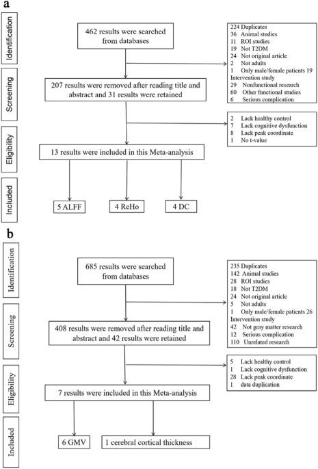
Experimental
Thirty healthy male Albino Wistar rats (180–200 gm) were maintained under controlled conditions (23 ± 2 °C, 55 ± 5%: humidity, 12 h L/D cycle). Institutional Animal Ethics Committee of Amity University Lucknow Campus approved the protocol (AUUP/AIP/4.2/2021). All animals were categorised into five groups as Normal control, Negative control, Geraniol 200 and 400 mg/kg and Standard (Atorvastatin 5 mg/kg) group. Obesity was induced with the administration of HFD (31% fat, 12% protein, 46% carbohydrate; 516.5 Kcal/100 g of feed) for the duration of four weeks excluding the normal control group [14]. At the end of fourth week blood glucose was measured by glucose oxidase peroxidase (GOD-POD) method in all the groups. Geraniol treated groups received geraniol 200 and 400 mg/kg p.o. [15] and Standard group received atorvastatin (20 mg/kg BW p.o.) for the duration of four weeks from fifth to eighth week. Blood glucose was estimated in all the groups by GOD-POD method at the end of fourth and eighth week of protocol. Feed intake was recorded daily, and body weight was monitored every week during complete protocol. Rats were fasted overnight for the estimation of OGTT at the end of protocol.
Collection and preparation of blood sample
Blood was withdrawn through the retro orbital for blood glucose measurement. Further prior the day of sacrifice, the rats were fasted overnight, and the blood was collected from the lateral tail vein by puncturing with needle for estimation of oral glucose tolerance test (OGTT). Serum samples were collected from blood using a centrifuge operated at 3000 rpm for 10 min and stored in the refrigerator at 4 °C prior to biochemical analysis (blood glucose and lipid profile).
Isolation of adipose tissue
The rats were sacrificed by cervical dislocation after eight weeks and restrained on dissection tray placing the dorsal side of body upward. The skin of rat was held up near the neck region and an incision was made with the help of scissor. The upper layer of skin was then cut open with the help of scapula and forceps. A butterfly shaped region of fat was exposed which was isolated to obtain the interscapular WAT and BAT. The rat was then pinned on the dissection tray facing the ventral side upward. Upper layer of skin was held up and incision was again made on the abdominal region to cut open the upper layer of skin by scrapping the sub cutaneous layer to obtain the sub cutaneous fat. The epididymal, inguinal, mesenteric, triceps, intraperitoneal adipose tissues were located and isolated by scrapping or by cutting out properly.
Isolation of liver, pancreas and heart
The abdomen was cut open to the thoracic region with the help of scissor and organs were kept apart with the help of forceps. Diaphragm was punctured with scissor and pulled it away from ribs. Heart was isolated by making an incision using fine scissors. Ribs were cut from both sides and lung was removed with fine scissors so that the heart was exposed. Heart was pinched downward using forceps and heart tissue was perfused with cool phosphate until no more blood is visible in the coronary arteries. Then after liver and pancreas were pulled out of abdominal cavity. Liver was separated from diaphragm by cutting the falciform and coronary ligament that attached the liver to diaphragm. All these tissues were excised and washed in cold phosphate buffer saline (PBS) pH 7.4. The tissues were then dried in folds of tissue paper and weighed.
Assessment of oxidative stress parameters
Oxidative stress parameters like MDA and SOD level were estimated in the homogenate of liver tissue as per previously reported methods [16]. Briefly, 2.5 mL Methionine, 0.3 mL Riboflavin, 0.1 mL NBT, 0.1 mL liver homogenate were uniformly illuminated with incandescent light for 15 min and level of SOD was estimated by determining absorbance at 560 nm. Moreover, lipid peroxidation was estimated by determine level of MDA with thiobarbituric acid at 532 nm wavelength.
Histopathological study
The pancreas, heart, and adipose tissue were isolated, flushed with ice-cold PBS, and fixed in 10% neutral buffered formalin while the heart was fixed in 4% formalin. Using a microtome, tissue was paraffin embedded and cut into pieces 5 mm thickness. The sections were placed on glass slides coated with poly L-lysine and stained using H&E staining with a Nikon Eclipse 80i microscope (Nikon, Kawasaki, Japan), the slides were examined after being photomicrographed in three randomly selected fields from each slide.
In silico
PubChem compound database (https://pubchem.ncbi.nlm.nih.gov/) was used to take the structure of geraniol in SDF format, which was converted with OpenBabel in to PDB format, which was further stored as pdbqt file, used for docking study. Protein Data Bank (PDB) utilized to retrieve the 3D X-ray crystal structure protein having 1HWK PDB code. Docking study was performed with Docking Autodock 4.2 software, protein was cleared from pdb chain A with Discovery Studio Visualizer v20.1.0.19295 software by removing Hetatms. Later, autodock 4.2 software was utilized to open the protein file, kollman charges and computing of Gasteiger charges were added after the addition of polar-H atoms. Macromolecule file was stored as Pdbqt file after merged all the non polar-H atom with autodock tool, this file was used for further study.
Active centres of protein were estimated with Autodock 4.2 and geraniol file was used as ligand, globally optimized conformation was identified with Lamarckian genetic search algorithm (LGA). The other parameters for docking calculation used were population size, 150; mutation rate, 0.02; and cross-over rate of 0.8; 2.5 × 10⁵ operations were executed for the generation of new docking trail. Grid box was decided on the target protein with X, Y and Z coordinates. Ligand, a grid spacing of 0.375 Å was used around the docking area and binding energy of geraniol with protein was analysed with Autodock 4.2, which was used for the estimation of hydrophobic interaction and H-bonds between target protein and ligand.
Statistical analysis
Data are expressed as mean ± SEM (n = 6). One-way analysis of variance (ANOVA) followed by post-hoc Dunnett test was used to compare the various groups. Two-way repetitive measure ANOVA followed by post-hoc Bonferroni test was used to find the differences in MWM acquisition data. p < 0.05 was considered statistically significant.


Comments (0)