Screening for Acrylamide-Degrading Fungi
Twenty-five fungal isolates from our laboratory stock [23][24,25,26,27,28] were grown on modified Potato Dextrose Agar (PDA) media (250-g potato extract, 2-g acrylamide, and 20-g agar–agar dissolved in 1 L distilled water) and incubated for 8 days at 30 °C. Among the experimented fungi, four fungal isolates mainly Aspergillus fumigatus, A. flavus, A. niger, and A. awomari had a reliable visual growth on the acrylamide containing PDA media. The isolates Aspergillus fumigatus, A. flavus, and A. niger were originally recovered from the soil samples from the Botanical Garden of Faculty of Science, Zagazig University, Zagazig, Egypt, in March/2021. Different acrylamide concentrations (0.01, 0.5, and 1.0%) were amended to the modified PDA media, the fungal culture was incubated at 30 °C for 10 days, and the fungal growth was observed, as well as modified Potato Dextrose Broth (PDB) media (250 g potato extract/ liter distilled water) were prepared, amended with different concentrations of acrylamide (0.01, 0.5 and 1.0%) and then inoculated with a plug of 7-day-old fungal culture, per 250-ml Erlenmeyer conical flaks. The cultures were incubated for 10 days at 30 °C, and the fungal biomass was collected, washed, and then assessed [29].
Morphological and Molecular Identification of the Potent Acrylamide-Degrading Fungi
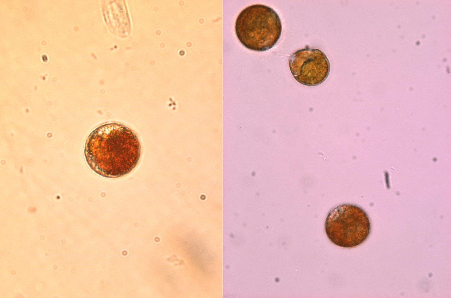
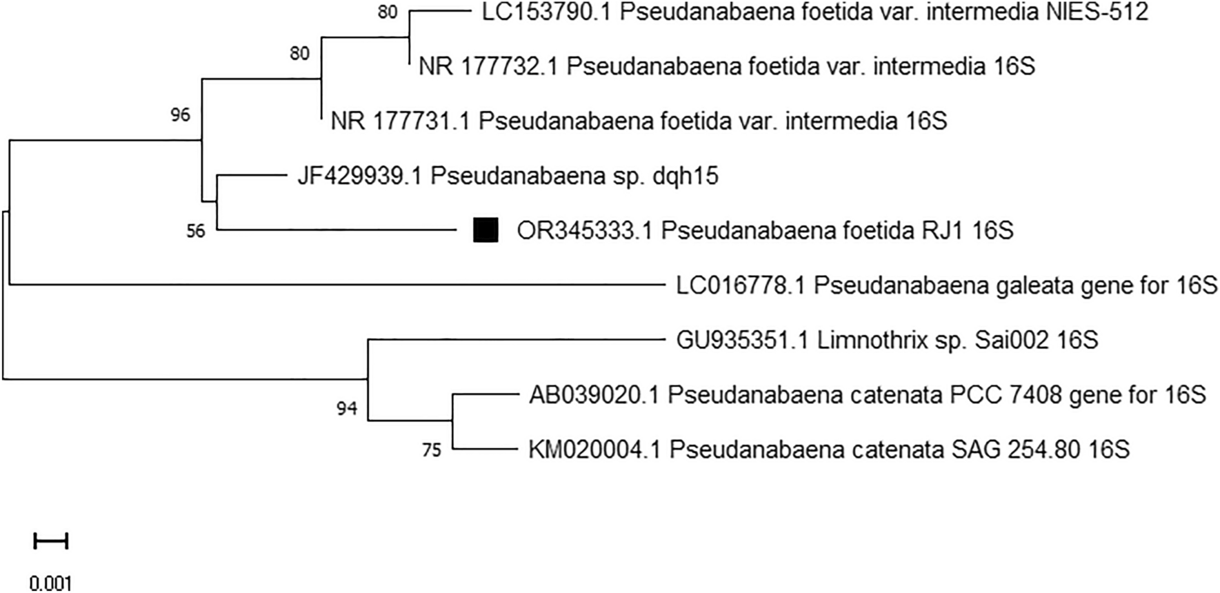
The experimental fungal isolates were identified based on their macroscopic and microscopic features by growing on PDA and Czapek-Dox media, according to the reference keys [30,31,32]. The most potent fungal isolate-degrading acrylamide was further molecularly confirmed based on its internal transcribed spacer (ITS) sequence [33,34,35]. The fungal mycelia (~ 0.2 g) were pulverized in liquid nitrogen and dispensed in 1-ml CTAB extraction buffer (2% CTAB, 2% PVP40, 0.2% 2-mercaptoethanol, 20-mM EDTA, 1.4-M NaCl in 100-mM Tris − HCl, pH 8.0). The genomic fungal DNA was used as template for PCR, with the primer sets ITS4 5′-GGAAGTAAAAGTCGTAACAAGG-3′ and ITS5 5′-TCCTCCGCTTATTGATATGC-3′. The reaction mixture contains 10-μl 2 × PCR master mixture (Cat. No. 25027), 1-μl gDNA, 1 μl of each primer (10 pmol/μl), and 20 μl distilled water. The PCR conditions were programmed as follows: initial denaturation 94 °C for 4 min, denaturation at 94 °C for 30 s, annealing at 53 °C for 20 s, extension at 72 °C for 40 s for 35 cycles, and final extension at 72 °C for 5 min. The amplicons were analyzed by 1.5% agarose gel in 1 × TBE buffer (Cat# AM9864) with 1-kb DNA ladder (Cat. #PG010-55DI). The amplicon was sequenced by Applied Biosystems, HiSQV Bases with the same primers. The sequence was annotated by non-redundant BLAST search on NCBI database, aligned with Clustal W and the phylogenetic tree was constructed with neighbor-joining method of MEGA X [36].
Acrylamide Amidase Activity and Protein Concentrations Assay
The most potent acrylamide-decomposing fungal isolate was grown on potato dextrose broth medium with 0.5% acrylamide for 10 days at 30 °C, then the fungal biomass was collected by filtration, and then intracellular crude proteins were extracted [18, 37,38,39]. Briefly, ten grams of the fungal biomass were pulverized in liquid nitrogen and then dispensed in 50-ml potassium phosphate buffer (pH 7.0) with 1-mM dithiothreitol and 1-mM EDTA. The mixture was vortexed for 5 min, centrifuged at 8000 rpm for 10 min at 4 °C, and the supernatant was used as a crude source for acrylamide amidase. The activity of acrylamide amidase was determined by Nessler’s reagent [19]. The reaction mixture contains 50-mM acrylamide in 20-mM potassium phosphate buffer (pH 7.0) and 500 μl of enzyme preparation in 1-ml total volume, the reaction was incubated at 40 °C for 15 min, stopped by adding 10% Trichloroacetic acid, and then the supernatant was amended with Nessler’s reagent (ADWIC, N0178111) [38, 39]. Blanks of the crude enzyme extract without acrylamide and substrate without enzyme, amended with Nessler’s reagent, were used. The developed color was measured at λ425 nm, and the concentration of ammonia was calculated from the inference of authentic concentrations of ammonium sulfate [19, 24, 25]. The activity of amidase (1 unit) was expressed by the amount of enzyme releasing 1 μmol of ammonia per mg protein under the standard conditions. The enzyme protein concentration was measured by Folin’s reagent [40], regarding to bovine serum albumin as authentic one. The activities and concentrations of L-asparaginase were assessed.
Purification and Molecular Subunit Structure of Aspergillus fumigatus Acrylamide Amidase
Aspergillus fumigatus was grown on potato dextrose broth medium amended with 0.5% acrylamide, incubated at the desired conditions, the fungal pellets were collected, and then washed by potassium phosphate buffer. The fungal biomass was pulverized in liquid nitrogen and the intracellular crude proteins were extracted [18, 38, 39, 41]. The crude enzyme was fractionally concentrated with 20-kDa cut-off dialyzer (Cat.# 546–00051), against polyethylene glycol 6000 [24, 26,27,28, 37, 38] followed by 30-kDa Ultracentrifuge membrane (Amicon, Millipore) at 10,000 rpm for 15 min at 4 °C. The enzyme was further purified by gel-filtration with Sephadex G200 column [33,34,35, 39]. The activity of amidase was measured by the standard assay, and the most active fractions were collected, concentrated, and their homogeneity was checked by denaturing PAGE [42]. The most active fractions were collected, further concentrated by 30-kDa Ultracentrifuge membrane, and further purified by ion-exchange chromatography with DEAE-Sepharose column [36,37,38]. The most active amidase and molecularly homogeneous fractions were selected. The molecular homogeneity and subunit structure of the purified acrylamide amidase were checked by SDS-PAGE [42], normalizing to authentic protein marker (Puregene, Cat. #PG-PMT2962, 315–10 kDa).
Biochemical Properties of the A. fumigatus Acrylamide Amidase
The biochemical properties of the purified A. fumigatus amidase such as reaction temperature, reaction pH, and thermal stability were investigated [19]. The reaction mixture containing 50-mM acrylamide dissolved in 20-mM Tris–HCl (pH 8–9), potassium phosphate buffer (5–7), and citrate phosphate buffer (4–7) was incubated at 40 °C for 15 min and then the enzyme activity was measured, as mentioned above. The standard reaction mixture was incubated at various temperatures (10 to 60 °C) and then the enzymatic activity was measured as mentioned above. The thermal stability of the purified amidase was assessed by pre-incubating the enzyme at 4, 20, 30, 40, and 50 °C for 30, 60, and 120 min and then measuring the residual enzyme activity as described above [18, 37, 43,44,45]. The impact of different cations on enzyme activity was determined by pre-incubating the apo-enzyme with different inhibitors (Ba2+, Fe3+, Ca2+, Hg2+, Fe3+, Al3+, Zn2+, Na+, Cu2+) for 2 h at 4 °C at final concentration 1 mM and then assessing the residual enzymatic activity. Different amino acid analogues such as 5,5′-dithio-bis-(2-nitrobenzoic acid) (DTNB), hydroxylamine, guanidine thiocyanate, iodoacetate, 3-methyl-2-benzo-thiazolinone hydrazone (MBTH), and phenylmethylsulfonyl fluoride (PMSF) were incubated with the enzyme at 1 mM for 2 h at 4 °C and then measured their residual enzyme activity.
Food Applications of the Purified A. fumigatus Amidase in Different Food Products
The functionality of amidase to degrade acrylamide in different food products mainly meat, bread, cookies, and potato chips was determined [14]. One gram of the tested food products was soaked in 5 ml of the amidase preparations (85 μmol/mg/min) for 60 min at 40 °C, using distilled water as negative control. The food products were vigorously homogenized, and the homogenate was centrifuged at 8000 for 10 min and the amount of acrylamide was determined by HPLC (YOUNG In, Chromass, Korea) of reverse phase C18 column (Cat.# 959,963–902). The mobile phase was methanol/acetonitrile/water (90:5:5, v/v/v) at a flow rate 1 ml/min for 25 min, and the absorbance of acrylamide was measured at λ203 nm [14], compared to the authentic one (Cat. #. 79–06-1). The purity and concentration of the acrylamide in samples were determined from the retention time and peak area, normalizing to authentic one at λ203.
LC–MS Analysis of Acrylamide and its Derivatives in Food Products
The prepared homogenates of the food products were defatted with hexane, evaporated, and the concentration of acrylamide and its degradation by-products were determined [18]. The acrylamide and acrylic acid concentrations were determined using LC–MS (Waters Corp., Milford, MA01757, USA). The ESI–MS-positive ion acquisition mode was carried out on a XEVO TQD triple-quadruple instrument, with column ACQUITY UPLC-BEH C18 (1.7 µm, 2.1 × 50 mm), with solvent system of acetonitrile (A) and 0.1% formic acid (B). The elution was carried out at 25 °C with a flow of 0.2 ml/min, using the following gradients: at the beginning (10% A and 90% B); up to 10 min (increase of solvent A to 90% and decrease of solvent B to 10%); from 10 to 15 min (90% A and 10% B); and from 15 min to the end (10% A and 90%). The chemical identity of the acrylamide and acrylic acids was determined reliant of their mass spectra and retention time referencing to NIST and WILEY libraries.
Fungal Deposition
Aspergillus fumigatus EFBL has been deposited to the GenBank with accession # MW737636.1 and at Assiut University Mycological Center, Egypt, with deposition # AUMC14078.
Statistical Analysis
The experiments were performed in triplicates, and the results were expressed by mean ± STDV. The statistical analysis was assessed using one-way ANOVA (analysis of variance, SPSS software v.18) test, and the means were compared with Duncan’s test at 0.05 level.

留言 (0)