
Remember me


A literature search was carried out in PubMed for the period between January 1st, 2000 and August 30th, 2023. The syntaxes used were “endometriosis AND matched AND (art OR assisted reproductive technology OR IVF OR in vitro fertilization OR ICSI)” (98 papers retrieved) and “endometrioma AND (unilateral OR contralateral) AND (art OR assisted Reproductive technology OR IVF OR in vitro fertilization OR ICSI)” (94 papers retrieved). Only studies providing reliable and unbiased information on specific steps of the IVF procedure were considered. Reviews were cited if deemed useful. No efforts were performed to identify abstracts submitted to meetings.
Endometriosis and ovarian response to gonadotropin stimulationAccording to the meta-analysis by Hamdan and co-workers, which included 17 studies for a total of n = 17,593 IVF cycles, a lower mean number of oocytes retrieved per cycle was demonstrated in women with endometriosis compared to controls (mean difference: − 2.0, 95% CI: − 2.9 to − 1.1) [8]. One is tempted to speculate that endometriosis per se may reduce the number of oocytes retrieved.
Notably, when assessing the endometriosis-related influence on ovarian response, some confounding factors come into play, including: (i) prior surgery, which can affect ovarian reserve and responsiveness to stimulation; (ii) the incompleteness of oocyte retrieval. Regarding this latter point, physicians are generally concerned by the risk of endometrioma infection during oocytes retrieval and tend to avoid endometrioma transfixion. Moreover, due to endometriosis, ovaries may be dislocated in the pelvis, making the retrieval more difficult (Fig. 1) [12]. Accordingly, the frequency of incomplete follicular aspiration was found to be over three times more common in affected women [27].
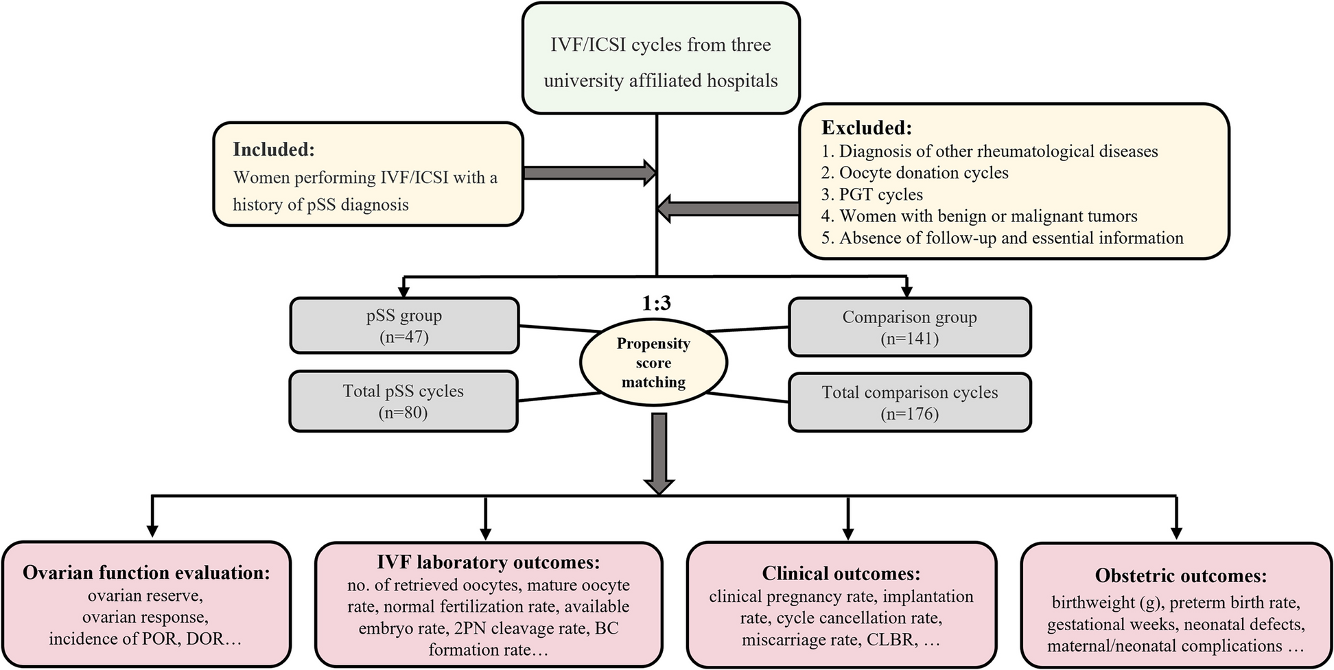
Insights from a rigorous matching designTo provide an unbiased evaluation of ovarian responsiveness in women with endometriosis, we have designed a study where n = 248 women with endometriosis and an adequate ovarian reserve (AMH > 1.1 ng/ml) were meticulously matched to n = 248 controls, according to age, pharmacological regimen (same drug, same initial dose), AMH concentration and study period [23]. Prior surgery for endometriosis or the presence of ovarian endometriomas were not exclusion criteria. This study design aimed to furnish an unbiased understanding of endometriosis’s effect on ovarian response. To concomitantly assess quantitative and qualitative aspects of the ovarian response, our primary outcome was the unavailability of good quality embryos on day 3 (not pregnancy rates as this might be influenced by the concomitant presence of adenomyosis). The rate of unexpected poor response (retrieval of ≤ 3 oocytes) according to the Poseidon Group (2016) as well as the overall success rate were secondary outcomes [28]. Results obtained showed that the number of women without good quality embryos did not differ between women with and without endometriosis (16% in both groups). However, in women with endometriosis, the duration of stimulation was longer, and the number of oocytes retrieved (but not mature oocytes) was lower. The rate of unexpected poor response to ovarian stimulation differed being 13% in non-affected cases versus 23% in controls (p = 0.005). Notably, in subgroup analyses, such higher rate of unexpected poor responders persisted only in women who had undergone surgery for the disease. All other variables related to ovarian response showed no notable difference (results are presented in Fig. 2).
Fig. 2
Box and whiskers plot of the number of follicles, oocytes retrieved, suitable oocytes, 2PN (fertilized oocytes), cleavage embryos and good quality embryos. Data from women with and without endometriosis are represented in red and green, respectively. A statistically significant difference emerged only for the number of oocytes retrieved (highlighted with an asterisk)
Albeit being a secondary outcome, it is worth noting that the cumulative clinical pregnancy and live birth rates were almost identical, even slightly favouring the endometriosis group (50% and 40% in endometriosis patients, and 49% and 36% in controls, respectively). Taken together, results from this study suggest that endometriosis per se does not have a major impact on folliculogenesis. The observed detrimental effect of surgery on the risk of unexpected poor response may reflect an increased difficulty in the oocyte retrieval procedure.
Another matched study published in 2017 should also be mentioned, although the sample size was smaller and the matching less scrupulous [29]. The authors retrospectively matched n = 119 women who had undergone surgery for endometriosis to a control group of n = 119 women without the disease by age, serum AMH, number of previous cycles and method of fertilization (conventional IVF or ICSI). The number of oocytes retrieved, and the number of good quality embryos were comparable. The live birth rate per cycle was also similar (27% vs 30%) [29].
The impact of ovarian endometriomasThe impact of endometriomas on ovarian response represents a related but independent issue. Several intra-patient comparisons between the two gonads (affected versus unaffected) have been performed to determine if unilateral ovarian endometriomas could affect ovarian response in women on ART cycles who had not previously had ovarian surgery [8, 12, 30,31,32,33]. These studies generally suggest that the presence of these cysts does not significantly impact ovarian response. Only one of these studies was prospective and reported also data on oocytes quality [34]. The number of developed follicles and oocytes retrieved were similar, being 3.7 ± 2.4 and 4.1 ± 1.7, and 4.2 ± 3.1 and 4.7 ± 2.5, respectively in the two ovaries. Fertilization and cleavage rates, and rate of high-quality embryos did not differ, being 64% and 64%, 58% and 51%, and 31% and 21%, respectively. However, the limited sample size (n = 29) and the small mean diameter of the endometriomas (25 ± 9 mm) hindered strong conclusions.
For bilateral endometriomas, three retrospective studies could be mentioned, of which one was very small (only n = 13 women) and not matched [35]. The second study, from our group, included n = 39 cases and n = 78 controls matched in a 1:2 ratio for age and study period [36]. Despite similar biomarkers of ovarian reserve, the number of follicles > 15 mm and oocytes retrieved were fewer in women with bilateral endometriomas compared to controls, being 6.2 ± 2.6 and 9.6 ± 4.8 (p < 0.001) and 7.1 ± 3.2 and 9.8 ± 5.5 (p = 0.001), respectively [36]. However, the cumulative live birth rate did not significantly differ, being 25% and 31%, respectively [36]. A third matched study enrolling n = 70 women with unoperated endometriomas, of whom n = 38 had bilateral cysts, failed to show any significant difference in serum AMH levels or number of embryos obtained. Notably, a subgroup analysis specifically focusing on these n = 38 women with bilateral endometriomas and their n = 38 matched controls was not reported [37].
A neglected but crucial aspect that could explain these inconsistencies is the size of the endometrioma. Several studies that examined the intra-patient comparison of ovarian response among women with unilateral endometriomas presented subgroup secondary analyses based on cyst diameter, suggesting a detrimental effect based on the cyst dimension [22, 38]. In general, firm conclusion could not be drawn because of the insufficient number of large endometriomas included and the nature of these analyses being secondary or exploratory. Ferrero et al. (2017) were the first to selectively focus on women with unilateral endometriomas larger than 5 cm. The intra-patient comparison showed a significant decline in ovarian response with a lower number of follicles in ovaries with endometriomas (2.6 ± 1.3) compared to healthy ovaries (4.8 ± 2.0; p < 0.001). Since the number of oocytes retrieved was recorded separately for the two ovaries, they were also able to report a marked difference between the affected and unaffected ovaries, which was 1.5 ± 1.1 and 3.3 ± 1.5, respectively (p < 0.001) [31].
A multicenter international study was then set aiming to identify the threshold of diameter above which ovarian response starts to be critically impaired [32]. The authors retrospectively included unoperated women carrying unilateral endometriomas with a mean diameter between 20 and 49 mm, and categorized them based on endometrioma size: 20–29 mm, 30–39 mm, and 40–49 mm. A negative effect on the number of developing follicles was observed only for cysts with a mean diameter from 40 to 49 mm. The median [interquartile range – IQR] number of developed follicles was 5 [3,4,5,6,7] and 7 [4,5,6,7,8] in affected and not affected ovaries, respectively (p = 0.01). These results suggest that a threshold of 4 cm might be used to discriminate between cysts that do and do not affect ovarian responsiveness [32].
Finally, a rather popular but poorly investigated aspect is represented by the possibility that the potential detrimental effect of endometriomas on ovarian reserve and response to gonadotropin might be progressive over time. In other words, recently developed ovarian endometriomas might initially present little to no issues whereas long-lasting lesions might pose significant risks. The biological plausibility supporting this view stems from the fact that ovarian endometriomas contain a plethora of potentially toxic agents. The long-lasting diffusion of these substances into the ovarian stroma may progressively damage and diminish the primordial follicular pool [39]. However, from the clinical point of view, this issue is controversial. Kasapoglu and coauthors repeated AMH testing at 6 months apart in n = 40 women with endometriomas (mean diameter 46 ± 17 mm, bilateral in 9 subjects) and n = 40 controls. They observed a statistically significant reduction of 26% (95% CI: 11–55%) in the formers, but no significant changes in the controls [40]. In contrast, we set up a study to retrospectively weight this aspect in women with endometriomas (average diameter of 26 ± 8 mm), who underwent more than one cycle of ovarian stimulation at intervals of more than 6 months (median 11 months, IQR 8–14 months). The contribution of the affected ovary to the overall response in terms of number of follicles retrieved remained consistent across cycles and equal to 44% (31–58%) during the first cycle and 44% (35–55%) in subsequent cycles [41]. From these two studies, we may infer that while the detrimental effects of endometriomas over time is unremarkable for small cysts, it could be significant for larger cysts.
Endometriosis and levels of steroid hormonesAccording to Barnhart and coauthors, women with endometriosis have a 19% reduction of peripheral of estrogen levels at the time of ovulation trigger [7], suggesting an altered steroidogenesis. Some molecular studies support a negative influence of endometriosis on growth, steroidogenic activity, and function of granulosa cells [42]. In affected women, both granulosa cell expression of P450 aromatase (an enzyme that converts androgens to estrogen) and estrogen concentrations in the granulosa cell culture mediums were found to be reduced [43].
However, when interpreting these findings, one cannot exclude a confounding effect arising from reduced ovarian reserve, at least when addressing evidence from clinical studies. The above-mentioned study from Invernici and co-authors (2020), who carefully matched cases and controls for ovarian reserve, tends to reject the hypothesis of perturbed folliculogenesis. The serum estradiol at the time of trigger was identical, the median [IQR] being 1837 [1283–2831] and 1901 [1341–2811] pg/ml in cases and controls, respectively [23]. Reschini and co-authors (2020) designed a study specifically tailored to address this issue. Matching n=53 cases and n=53 controls by study period, age, total number of developed follicles, protocol of ovarian stimulation, type and starting dose of gonadotropin, they reported similar median [IQR] serum estrogens of 1586 [1146–2787] and 1625 [1060–2322] pg/ml, respectively [26]. Overall, available clinical evidence challenges the data from basic science studies [42]. Ovarian steroidogenesis does not seem to be affected in women with endometriosis, further supporting the idea that the disease might have minimal, if any, impact on oocyte quality.
Endometriosis and fertilization rateAlthough the number of studies included was very limited, some meta-analyses reported a reduced fertilization rate per oocyte in women with endometriosis [4, 5]. According to Horton and co-workers (2019), this finding is significant for treated patients (OR 0.92, 95% CI: 0.86–0.99, p = 0.03) but not for those untreated [5]. Fertilization rate seems to be more compromised in case of milder endometriosis presentations. Though, the estimation of the fertilization rate in affected cases is as well not devoid of confounding factors.
Previous studies have retrospectively compared results between ICSI and conventional IVF (c-IVF) in women with endometriosis [43]. This was based on the assumption that endometriosis itself might be responsible for a reduced oocyte competence so that ICSI, rather than c-IVF, could overcome this oocyte impairment. Comparing sibling oocytes, Komsky-Elbaz et al. have reported a higher fertilization rate when ICSI was preferred rather than c-IVF in couples with stages III–IV endometriosis [43]. However, possible biases in the analysis should be kept in mind, including: (i) maturity of oocytes is routinely established in case of ICSI and this selection bias may contribute to a higher fertilization rate per oocyte compared with unselected oocytes undergoing c-IVF; (ii) the common tendence to prefer ICSI in cases of male infertility but also to avoid total fertilization failure.
A more accurate approach for the correct assessment of this parameter would be a comparison of the fertilization rate of oocytes from women with and without endometriosis by means of the same insemination approach. Along this line, the above-mentioned study from Shebl et al. is of great interest because the authors ensured matching based on the fertilization procedure used [29]. They observed comparable fertilization rates for women requiring ICSI and a slightly lower rate among those endometriosis women treated with c-IVF (45% versus 54%, p = 0.03). Again, a potential bias could be introduced as the analysis was performed per oocyte (and not per woman). In a recent matched case–control study, we have demonstrated that a diagnosis of endometriosis does not negatively affect the performance of c-IVF [29]. Three-hundred and fourteen patients with endometriosis and normozoospermic partners have been matched in a 1:1 ratio with patients undergoing IVF for other indications, with respect to age (± 6 months), number of oocytes retrieved (± 1), and study period. The fertilization rates did not differ between women with and without endometriosis (median [IQR] being 78% [60–100%] and 75.0% [56–90%]; p = 0.24, respectively) [24]. A similar approach should be adopted for ICSI in endometriosis patients with also a male infertility factor to prove that the fertilization rate is not substantially compromised in women with endometriosis requiring ICSI. To date, it can be reasonably inferred that endometriosis does not impact the performance of c-IVF.
Endometriosis and embryo quality and ploidyThe assessment of embryo morphology and ploidy rate in women of endometriosis, as a measure to quantify the impact of the disease on ART outcomes, is not devoid of problems. Firstly, morphological features are characterized by differences in the criteria adopted to evaluate embryo quality, leading to inconsistencies across studies. Furthermore, both embryo morphology and ploidy seem to be at some extent affected by the ovarian reserve and the dose/duration of gonadotrophin regimen used for ovarian stimulation [44]. This leads to the idea that retrospective studies addressing this question may have been confounded by the possible inclusion of affected women who have undergone surgery. Despite these possible limitations and confounders, a recent meta-analysis based on 22 studies, specifically addressing high embryo quality rate as main outcome measure, did not show any negative impact of endometriosis [45]. Women with endometriosis, including severe stages and endometriomas had similar rates of embryo formation, cleavage embryos and high-quality embryos rates compared with the control group [45].
Sanchez et al. analysed n = 429 ART cycles in women undergoing surgery for moderate/severe stages and compared them with n = 851 cycles in control patients matched for age, number of oocytes retrieved and study period [46]. No differences were reported in terms of number of cleavage stage embryos and proportion of good/fair quality embryos. In contrast, this study documented a reduced likelihood of pregnancy in the endometriosis group, which may be explained by the higher doses of gonadotropins required in the endometriosis group to achieve the same number of oocytes [19, 20]. Furthermore, the conclusions of this study are limited by the inclusion of cycles adopting only cleavage stage embryo transfer strategy, the exclusion of cycles where no embryo was obtained or all embryos were cryopreserved, and by limited attention given to the selection of controls.
Going further, Vaiarelli and coworkers have evaluated the euploid blastocyst rate per cohort of inseminated metaphase II oocytes [47]. Affected patients (n = 210) were matched in a 1:2 ratio to controls (n = 420) by IVF clinic, maternal age at retrieval, number of previous failed IVF treatments and number of metaphase II oocytes retrieved. The blastocyst rate and the embryo euploid rate per cohort of fertilized oocytes was similar between cases and matched controls, even if the blastocyst morphology was not considered.
Only two other studies have examined the euploid rate of embryos from patients with endometriosis. Results are controversial. In 2017, Juneau et al. retrospectively analysed the aneuploidy rate of 1880 blastocysts obtained from patients with endometriosis and compared them with 23,054 blastocysts from age-matched controls. They reported similar aneuploidy rates per biopsied blastocyst in the two groups [48]. In disagreement, Yan and coworkers, evaluating 7092 biopsied embryos, found a lower euploid embryo rate in women with endometriomas compared to controls (53% vs. 62%, p = 0.012) [49]. However, in this latter study, the statistical differences between the two groups in terms of total and starting dose of gonadotrophins used and FSH levels, question its absence of confounding factors. In this regard, based on the study design employed, results from Vaiarelli and coworkers seem the most robust [47].
Endometriosis and embryo implantation rateEmbryo implantation potential is one of the most debated aspects of endometriosis-related infertility and IVF failure. An altered receptivity was advocated as a main reason for the lower pregnancy rate in women with endometriosis, beyond the lower ovarian reserve. A burden of literature has documented molecular and cellular alterations in the eutopic endometrium of women with endometriosis. These molecular pathways can be broadly classified into several groups including epigenetic modifiers, immune response regulators and inflammation triggers, hormonal stress inducers, epithelial-mesenchymal transition modulators [50]. Given these premises, it has been hypothesized that the communication between embryo and endometrium could be impaired, increasing the risk of implantation failure [51]. The inflammatory milieu of the pelvis has also been supposed to have some echoes in the endometrial cavity (secondary event). Regardless of the pathogenetic pathways leading to altered endometrium (i.e., whether they are primary or secondary of the disease, or both), ART is not the solution for these detrimental mechanisms. ART treatments can overcome most of the anatomic and functional impairment of the reproductive system, but they cannot heal the supposed molecular endometrial alterations.
Notably, measuring endometrial receptivity is the most challenging step in case of endometriosis. Embryo implantation is influenced by two main confounding factors. First, the low ovarian reserve reduces the rate of optimal embryos to transfer. In addition, poor responders are at higher risk of early progesterone elevation [52, 53], a condition that displace the window of implantation, therefore interfering with embryo implantation [54]. Second, endometriosis is associated with conditions that per se interfere with implantation, including adenomyosis, polyps and endometritis [3, 55,56,57]. Endometrial polyps and chronic endometritis are thought to exert a negative effect on endometrial receptivity [55, 57,58,59]. Adenomyosis is thought to prompt both uterine hyperperistalsis and fibrosis through epithelial-to-mesenchymal transition and fibroblast-to-myofibroblast transdifferentiation. Notably, the number of microvilli is reduced, steroid hormone metabolism is altered, and oxidative stress is increased in the endometrium of women with adenomyosis [3].
Clinical studies specifically designed to investigate the detrimental effect of the disease on endometrial receptivity are therefore difficult to conduct [59,60,61,62,63]. Analysing data derived from the ‘freeze all’ strategy could represent a way to eliminate some of the confounders. In a retrospective Chinese cohort study based on more than n=400 endometriosis patients undergoing frozen embryo transfer after ART treatments, affected patients were matched in a 1:3 rate with women undergoing ART due to tubal factor-related infertility, considering their age, infertility duration, serum FSH levels, antral follicular count, and BMI. Results obtained showed that endometriosis patients have lower live birth rate per transfer, as well as lower cumulative live birth rate, compared to controls [63]. However, the number of oocytes retrieved was significantly lower in affected women. As already discussed [46], this may affect the chance of pregnancy because embryos obtained with higher doses of gonadotropins or lower number of oocytes are at higher risk of aneuploidy. Accordingly, Blank et al. also observed a detrimental effect on pregnancy rates when comparing fresh transfers between women with and without endometriosis, after matching them for study period, age, parity, and embryo quality [62]. However, the number of retrieved oocytes were again significantly lower among women with endometriosis, as well as the rate of c-IVF (controls predominantly resorting to ICSI due to male factor infertility). Not surprisingly, other studies present differing views. Bishop et al. evaluated the implantation trend in three populations undergoing euploid frozen embryo transfer after ART treatments for different indications, including endometriosis, male factor, and preimplantation genetic testing for monosomic disorders. This study design overcomes the limitations of the three beforementioned studies. No difference in preg
Comments (0)